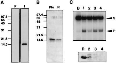
Figure 3.
Immunological identification of Hjc in P. furiosus cells. (A) The sonication extract of recombinant cells was separated by 15% SDS/PAGE, transferred onto PVDF membranes and treated with immune (I) or preimmune (P) sera raised against Hjc. (B) Fraction V from P. furiosus cells (Pfu) and 17 ng of recombinant Hjc protein (R) were analyzed by Western blotting with anti-Hjc serum. (C) Immunodepletion analysis. (Top) Endonuclease assay of each depleted extract. (Bottom) Each precipitant was analyzed by Western blotting with anti-Hjc serum. Lanes: B, buffer A; 1, crude extract before antiserum treatment; 2, anti-Hjc serum; 3, anti-Pfu-PCNA serum; 4, anti-PI-PfuI serum. Lane R was loaded with 15 ng of recombinant Hjc.

