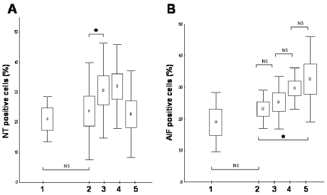
Figure 4.
Tyrosine nitration (A) and AIF translocation (B) determined by immunohistochemistry scores. After staining, NT-positive cells were counted in peripheral leukocyte smears. Results are expressed as mean (represented by squares) ± SEM (represented by boxes) and ± SD (represented by bars). Lane 1 indicates control samples from stable angina patients; lanes 2–5 show NT-positive cell counts or AIF translocation–positive cell counts in patients with acute myocardial infarction before coronarography (lane 2), immediately after the successful primary PCI (lane 3), 24 ± 4 h after reperfusion of the ischemic myocardium (lane 4), and 96 ± 4 h after PCI (lane 5). Primary PCI induced an immediate increase in tyrosine nitration, whereas a gradual increase of AIF translocation was observed at 24- and 96-h time points after reoxygenation of the ischemic myocardium. *P < 0.05; NS, nonsignificant.

