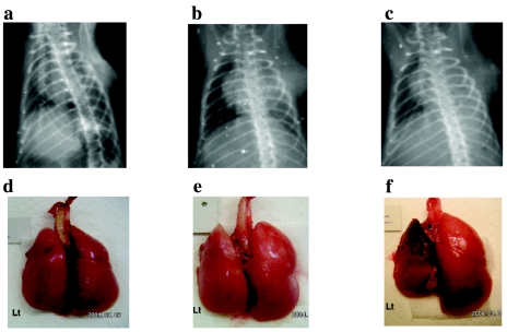
Figure 2.
Chest radiographic findings and macroscopic view of the allografts. Upper panel: Chest x-ray of allograft recipients on day 7 after transplantation. (a) Group A (antileukinate early administration) (AS = 6). (b) Group B (antileukinate continuous administration) (AS = 5). (c) Cont-B allograft recipient (AS = 1). Lower panel: Gross anatomy of group A allograft (d), group B allograft (e), and Cont-B allograft (f).

