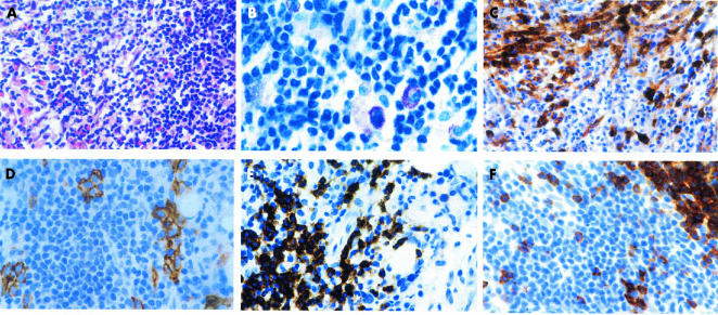
Figure 1.
Bone marrow involvement in indolent systemic mastocytosis; a 55 year old female patient with adult-type urticaria pigmentosa. (A) A focal compact bone marrow infiltrate containing spindle shaped mast cells, intermingled eosinophils, and a large accumulation of lymphocytes; haematoxylin and eosin stain. (B) A higher magnification reveals sheets of small to medium sized lymphocytes, with slightly pleomorphic nuclei. Note a few loosely scattered hypogranulated mast cells. A diagnosis of mastocytosis should not be made on the basis of such findings alone; Giemsa stain. (C) Immunostaining with an antibody against tryptase is clearly positive for the mast cell infiltrates. Note that a large proportion of the mast cells has a clear spindle shaped appearance; avidin–biotin complex method (ABC); anti-tryptase antibody (AA1). (D) Disseminated groups of mast cells with expression of CD25 can be easily detected within the large lymphoid infiltrate; ABC method; anti-CD25 antibody. (E) A large proportion of the lymphocytes expresses the B cell associated antigen CD20; ABC method; anti-CD20 antibody (L26). (F) T cells expressing the antigen CD2 occur in nearly equal numbers to B cells (see (E)). Note that a large cluster of lymphocytes remains unstained; ABC method; anti-CD2 antibody.

