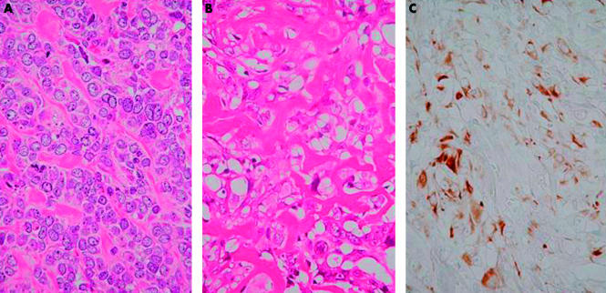
Figure 3.
Case 2. (A) Photomicrograph showing the epithelioid cytological features of the tumour cells—eccentrically located vesicular nuclei with prominent nucleoli and abundant palely eosinophilic cytoplasm (haematoxylin and eosin stained; original magnification, ×400). (B) Photomicrograph showing an area of lace-like osteoid formation (haematoxylin and eosin stained; original magnification, ×400). (C) Immunohistochemical stain for cytokeratin showing diffuse and intense positivity of tumour cells (original magnification, ×400).

