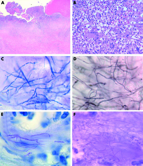
Figure 2.
(A) Low power magnification of the pleura (haematoxylin and eosin stain). (B) High power magnification of the granulomatous tissue, depicting the infiltration of giant cells, lymphocytes, and neutrophils (haematoxylin and eosin stain). (C) Gram staining revealed the thin microorganisms with a filamentous growth pattern. (D) The bacilli were positively stained with Grocott’s methenamine silver. (E) The bacilli exhibited weak acid fast reactivity by the Fite-Faraco staining method. (F) The microorganism exhibited only a weak reaction with the periodic acid Schiff stain.

