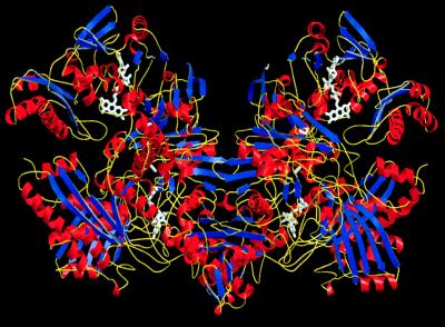
Figure 1.
Ribbon representation of the CO dehydrogenase dimer. The twofold-symmetry axis of the dimer runs vertically in the plane of the figure. Helices are shown in red, β-sheets in blue, and the connecting turns and loops in yellow. The cofactors are depicted in white. Structure presentations were created by using bobscript and raster3d.

