
Figure 2.
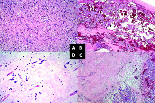
(A) Typical predominantly spindle cell gastrointestinal stromal tumour with heterogeneous nuclear atypicality and a mitotic activity averaging 18 mitoses/50 high power fields (original magnification, ×200). (B) Intratumorous haemorrhagia but no necrosis at the time of the first surgical intervention (original magnification,× 40), with (C) a decrease in cellular cohesion, shrinkage of the tumour tissue, and widespread haemorrhagia (original magnification, ×40). No mitoses were seen, and immunohistochemistry revealed a proliferative fraction of almost zero, as determined by staining for MIB-1. (D) The gelatinous changes to the paucicellular tumour tissue remained, and tumour tissue was still non-proliferating (original magnification, ×200). All slides were stained with haematoxylin and eosin.