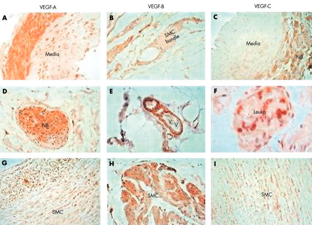
Figure 1.
Immunohistology for vascular endothelial growth factor (VEGF) isoforms. (A–C) Sections of uterine artery. (A) Staining for vascular endothelial growth factor A (VEGF-A): positive staining is localised predominantly to the adventitia, with sporadic staining of the media (original magnification, ×400). (B) Staining for VEGF-B: positive staining in smooth muscle cells (SMCs) within the collagenous tissue in the extracellular matrix (original magnification, ×400). (C) Staining for VEGF-C: localisation of VEGF-C to tissues in the adventitia (original magnification, ×200). (D–F) Sections of abdominal aortic aneurysm (AAA) tissue. (D) Localisation of VEGF-A to a nerve bundle (NB) (surrounded by the perineural sheath) within the adventitia (original magnification, ×400). (E) Localisation of VEGF-B to SMCs around a vasa vasorum (VV) in the adventitia (original magnification, ×400). (F) Localisation of VEGF-C to leucocytes within a vasa vasorum (original magnification, ×1000) but no staining of endothelium or SMCs. (G–I) Sections of carotid atheroma. (G) Localisation of VEGF-A to medial SMCs in diffusely thickened intima excised from an atherosclerotic artery (original magnification, ×200). (H) Localisation of VEGF-B to a bundle of SMC in the media of a complicated atheroma (original magnification, ×400). (I) Diffuse localisation of VEGF-C to the medial SMCs in an uncomplicated atheroma in a similar pattern to VEGF-A (original magnification, ×200).

