
Figure 4.
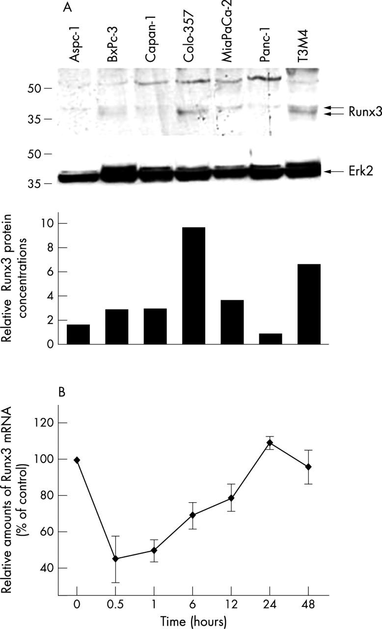
Runx3 expression and regulation in pancreatic cancer cell lines: (A) protein lysates (20 µg) of the indicated cell lines were subjected to Western blotting using a Runx3 antibody, as described in the Methods section (upper panel). Equal loading was determined by stripping the membrane and blotting with an antibody to Erk2 (middle panel). Relative amounts of Runx3 protein were determined by OD (Runx3)/OD (Erk2) × 100 for each sample (lower panel). (B) Colo-357 cells were incubated in the absence (0) or presence of 200pM transforming growth factor β1 for the indicated time. RNA was extracted and real time quantitative polymerase chain reaction for Runx3 was performed. The respective control mRNA expression values were set to 100%. Results are presented as mean ± SEM of three independent experiments. OD, optical density.