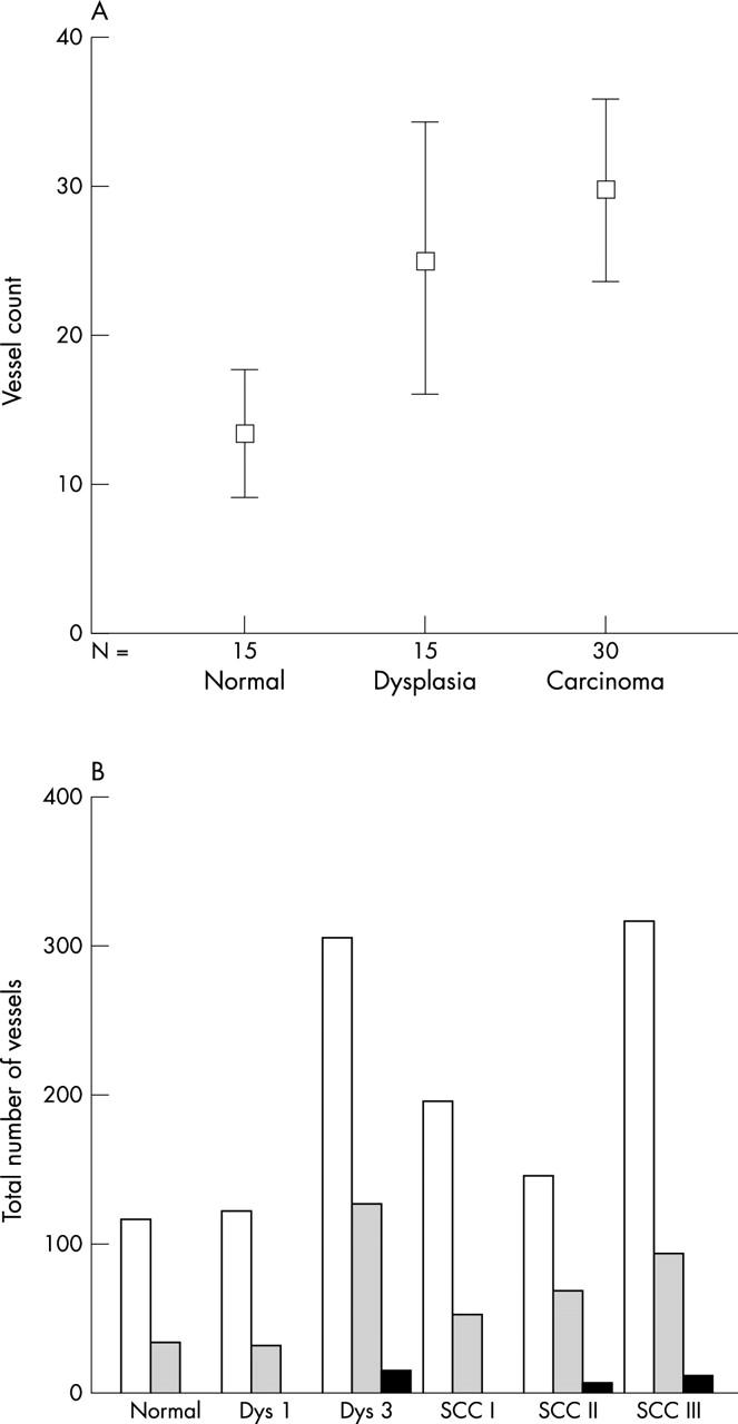
Figure 2.

(A) Automated quantitative image analysis of vessel count in each measurement area of factor 8 stained vessels in specimens of laryngeal normal tissue, dysplasia, and squamous cell carcinoma; means and 95% confidence intervals are shown. N, number of vessels analysed. (B) Total numbers of vessels in normal tissue, mild (Dys 1) and severe dysplasia (Dys 3), and squamous cell carcinoma (SCC) grades I, II, and III divided according to size. Open bars, 16–300 μm; shaded bars, 301–2800 μm; black bars, > 2801 μm.
