
Figure 1 .
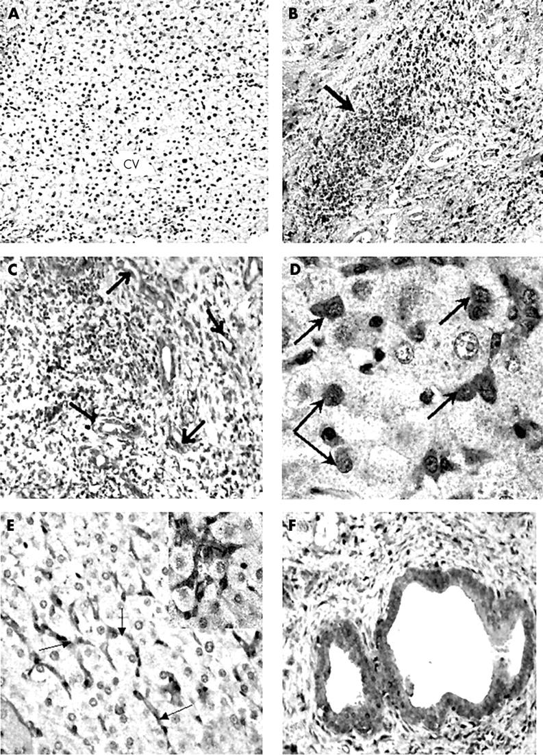
Immunoperoxidase showing COX-2 expression in (A) normal and (B–F) cirrhotic liver samples. (A) Normal human liver showing complete absence of COX-2 immunoreactivity. CV, central (terminal hepatic) vein region; original magnification, ×10. (B) A cirrhotic liver showing infiltration with inflammatory cells, which show dense COX-2 immunoreactivity (arrow); original magnification, ×25. (C) Large number of blood vessels seen in a cirrhotic liver showing COX-2 expression (arrows); original magnification, ×50. (D) Macrophage-like cells (arrows) infiltrating between hepatocytes show high COX-2 immunoreactivity; original magnification, ×100. (E) COX-2 immunoreactivity is seen in sinusoidal cells (arrows) in cirrhotic liver. The COX-2 positive sinusoidal cells are seen surrounding negatively stained hepatocytes; original magnification, ×50. (F) A liver cirrhosis sample showing COX-2 expression in the epithelial lining of bile ducts; original magnification, ×100.