
Figure 2 .
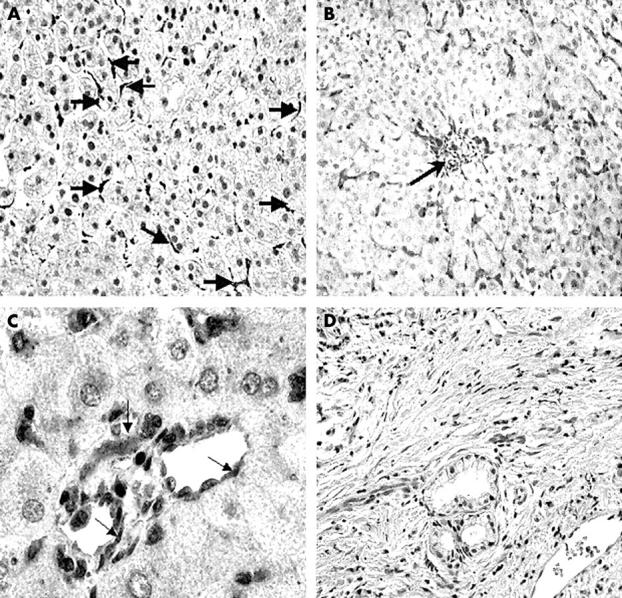
Immunoperoxidase staining showing COX-1 distribution in (A) normal liver and (B,C) human cirrhotic liver. (A) COX-1 is seen in normal liver, localised in the sinusoidal cells (arrows); original magnification, ×50. (B) In cirrhotic liver, COX-1 immunoreactivity is seen in the perivenular region (arrow) and in sinusoidal cells extending from the terminal hepatic vein; original magnification, ×25. (C) COX-1 is localised to the endothelial lining of two blood vessels seen in cirrhotic liver (arrows), with no expression in hepatocytes; original magnification, ×100. (D) A negative control in which the primary antibodies (COX-1 and COX-2) were omitted from the staining procedure, showing complete absence of staining and indicating the high specificity of the antibody used in our study; original magnification, ×50.