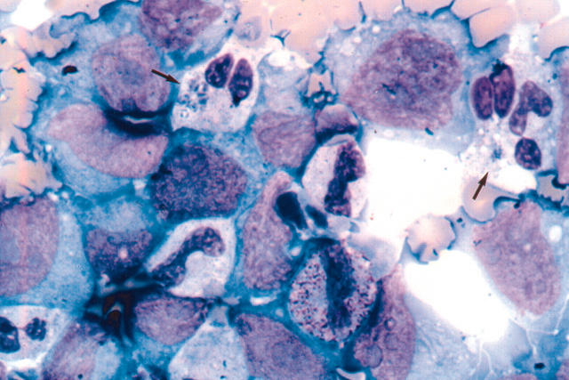
Figure 2.
Photomicrograph of cytospin preparation from culture aliquots of the patient’s blood obtained during the acute phase of human granulocytic ehrlichiosis after three days of culture incubation. Arrows show Anaplasma phagocytophilum inclusions in the patient’s granulocytes. Several morulae are present in the cytoplasm of the granulocyte indicated with an arrow at the top left hand part of the figure. Infection is not seen in the surrounding HL-60 cells.

