
Figure 4.
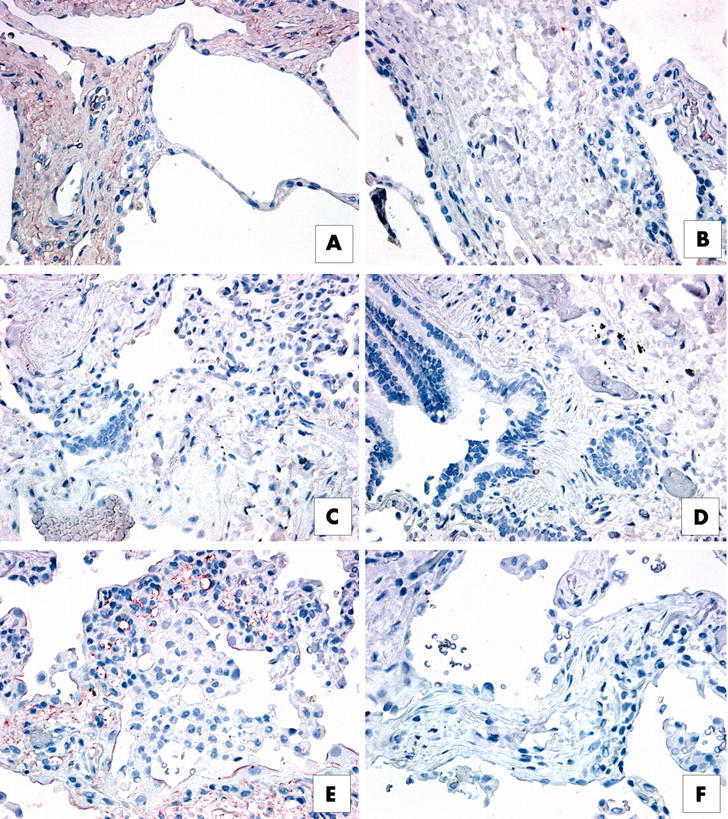
Immunohistochemical analysis of interleukin 13 receptor α1 (IL-13Rα1) in upper lobe surgical lung biopsies (SLBs) from patients with (A, B) respiratory bronchiolitic interstitial lung disease, (C, D) non-specific interstitial pneumonia (NSIP), and (E, F) usual interstitial pneumonia (UIP). Representative positive (red) staining for IL-13Rα1 is shown in panels (A), (C), and (E). Appropriate negative controls are shown in panels (B), (D), and (F). (A) IL-13Rα1 was expressed abundantly in the interstitium of SLBs from patients with RBILD. (B) IL-13Rα1 expression was associated with blood vessel walls in histological sections from patients with NSIP, whereas interstitial areas were lightly stained for this subunit. (E) IL-13Rα1 (red staining) was particularly prominent in pulmonary areas of neovascularisation in SLBs from patients with UIP.