
Figure 6.
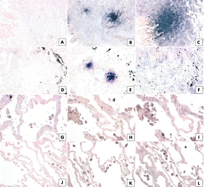
Immunohistochemical analysis of (B, E, H, K) interleukin 4 receptor α (IL-4Rα) and (C, F, I, L) IL-13Rα2 in upper lobe surgical lung biopsies (SLBs) from patients with (B, C) usual interstitial pneumonia (UIP), (E, F) non-specific interstitial pneumonia (NSIP), (H, I) respiratory bronchiolitic interstitial lung disease (RBILD), and (K, L) normal controls. Representative positive (purple) staining for IL-4Rα and IL-13Rα2 was detected in distinct foci in UIP SLBs (B, C), and IL-4Rα was also detected in distinct foci in NSIP SLBs (E). (H) Only mononuclear cells expressed IL-4Rα protein in upper SLBs from patients with RBILD. (I) Staining of mononuclear and interstitial cells for IL-13 Rα2 was seen in upper SLBs from patients with RBILD. (C) IL-13Rα2 was lightly stained in interstitial areas in SLBs from patients with NSIP. Rare IL-4Rα (K) and IL-13Rα2 (L) positive mononuclear cells were seen in SLBs from the normal controls. (A, D, G, J) Negative controls for each patient group. Black material (D, E, F) may be deposits as a result of cigarette smoke.