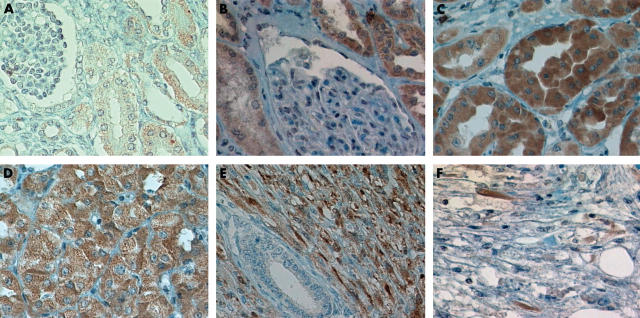
Figure 1.
Immunohistochemical expression of KIT. (A) Fetal and (B) adult renal tubules stained for KIT. Note the lack of staining in renal corpuscles. Diffuse intense staining for KIT in (C) conventional renal cell carcinoma, (D) oncocytoma, and (E) mesoblastic nephroma. (F) A few scattered spindle cells were positive for KIT in angiomyolipoma.

