Figure 2.

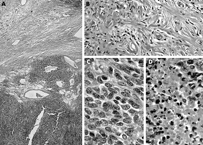
(A) Low power view of a cellular nodule and fibrous background. (B) The specimen had the classic appearance of a solitary fibrous tumour. (C) High power view of a cellular nodule. (D) Focal necrosis in a cellular nodule.

(A) Low power view of a cellular nodule and fibrous background. (B) The specimen had the classic appearance of a solitary fibrous tumour. (C) High power view of a cellular nodule. (D) Focal necrosis in a cellular nodule.