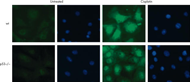
Figure 6.
Immunofluorescent detection of nuclear p73 in primary colonocytes using Alexa-488 conjugated secondary antibody. Nuclear accumulation of p73 is seen after treatment with cisplatin in both wild-type (wt) and p53 null cells using the sheep polyclonal Ab77 antibody. DAPI was used as a nuclear counterstain. The cells shown were exposed to cisplatin for 24 hours. Images were captured using a Hamamatsu chilled CCD camera and Zeiss fluorescent microscope.

