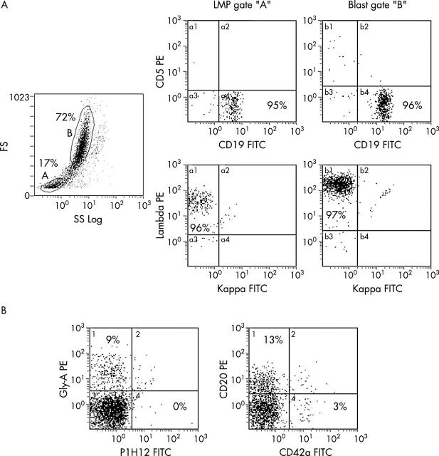
Figure 2.
Surface characteristics of leukaemia cell microparticles. (A) Surface immune phenotyping staining patterns in leukaemia/lymphoma cell microparticle (LMP) (gated as A) and leukaemic blast (gated as B) populations in a patient with Burkitt’s leukaemia. (B) Presence of LMPs (CD20 positive) along with platelets (CD42a positive) and erythrocytes (glycophorin A positive), but without endothelial cell particles (P1H12 positive) in a platelet poor plasma sample. The numbers on the histograms represent the percentage of events. FITC, fluorescein isothiocyanate; FS, forward scatter; PE, phycoerythrin; SS, side scatter.

