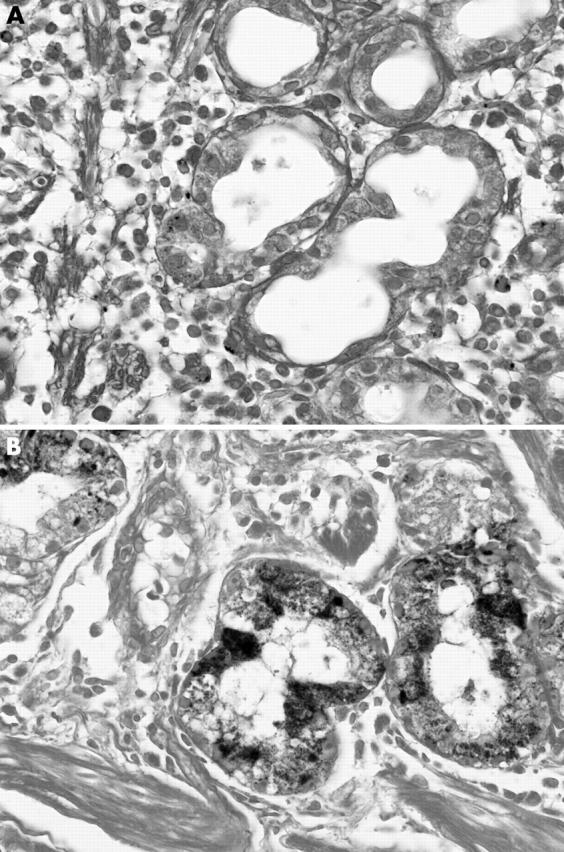
Figure 1.

Haemosiderin was seen in the cytoplasm of parenchymal cells of the gastric glands. (A) In this example of grade 1 deposition, both interstitial deposition and glandular deposition are scant. (B) In this example of grade 3 deposition, stainable iron is almost exclusively distributed within the glands (Perls’ stain).
