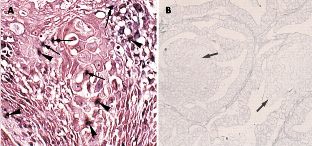
Figure 4.
(A) Demonstration of human papillomavirus (HPV) by the use of in situ hybridisation. HPV DNA (arrows and arrowheads) is demonstrated in the nuclei of cells with squamous differentiation in an endometrioid carcinoma. According to Cooper’s criteria, HPV was present in the integrated (arrowheads) and episomal (arrows) forms (type 3, Cooper’s criteria). Original magnification, ×150. (B) Demonstration of HPV DNA by use of in situ hybridisation. Neither the neoplastic glandular cells nor the morules harbour HPV DNA (endometrioid carcinoma case). The arrows indicate morules. Original magnification, ×150.

