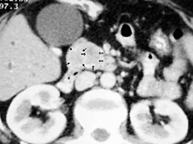
Figure 1.
Computed tomography (CT) showed a solid mass, located inferiorly to the pancreatic head and uncinate process. The mass enhanced similarly with pancreatic parenchyma at the early arterial phase. Preoperatively, the mass was interpreted as being located in the pancreatic head. CT images were evaluated retrospectively with the findings at surgery and two different lesions were differentiated (arrows).

