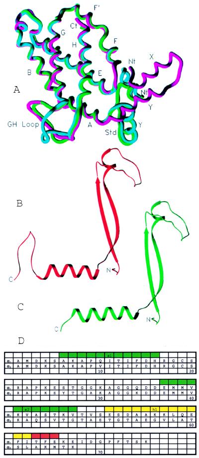
Figure 2.
Subunit secondary structures and sequences. (A) A superposition of β subunit C (blue), β subunit D (green) with the phycobilisome PE β subunit (red; ref. 10). Helices are labeled X, Y, A, B, E, F, F′, G, and H. All C termini are at one location, Ct, and the distinct N termini are labeled Nt. (B and C) Ribbon representations of the α1 (red) and α2 (green) subunits, respectively. (D) The aligned sequences of the α subunits along with the secondary structure. The green bars indicate β-strands 1 and 2. The yellow bar indicates the α-helix in the α1 chain. The helix in the α2 chain is longer by three residues (shown in red).

