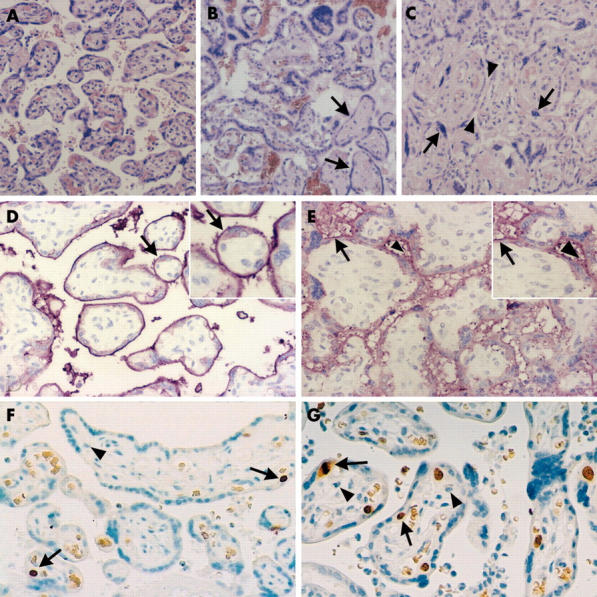
Figure 2.

Histopathological and immunohistochemical examination of the index and control placentas. (A–C) Haematoxylin and eosin staining of placental tissue sections (original magnification, ×100). (A) Normal term placenta, 38 weeks of gestation; (B) index placenta containing immature villi organised into proliferation centres and fibrotic, avascular villi (arrows); (C) villous crowding in the index placenta with pinched intervillous space (arrowheads) and syncytial knotting (arrows). (D, E) Immunohistochemical staining for alkaline phosphatase (AP) in placental tissue sections with inserts of higher magnification (original magnification, ×200). (D) In normal placentas, intensive linear positivity of the brush border membrane was detected (arrow), whereas in the index placenta, weak, diffuse, interrupted granular positivity of the membrane was seen (the arrow shows a normal part, whereas the arrowhead points to AP staining of the intervillous space). (F, G) Immunohistochemical staining for Ki-67 in placental tissue sections (original magnification, ×100). The nuclei of proliferating cytotrophoblasts were stained (one of the arrows shows a nucleus in the mitotic phase). (G) The ratio of proliferating cells in the index placenta was 8–10%, whereas it was approximately 1–2% in control placentas of the same gestational age (F). No Ki-67 staining was seen in either the syncytiotrophoblast layers or the villous stromal cells (arrowhead).
