
Figure 4.
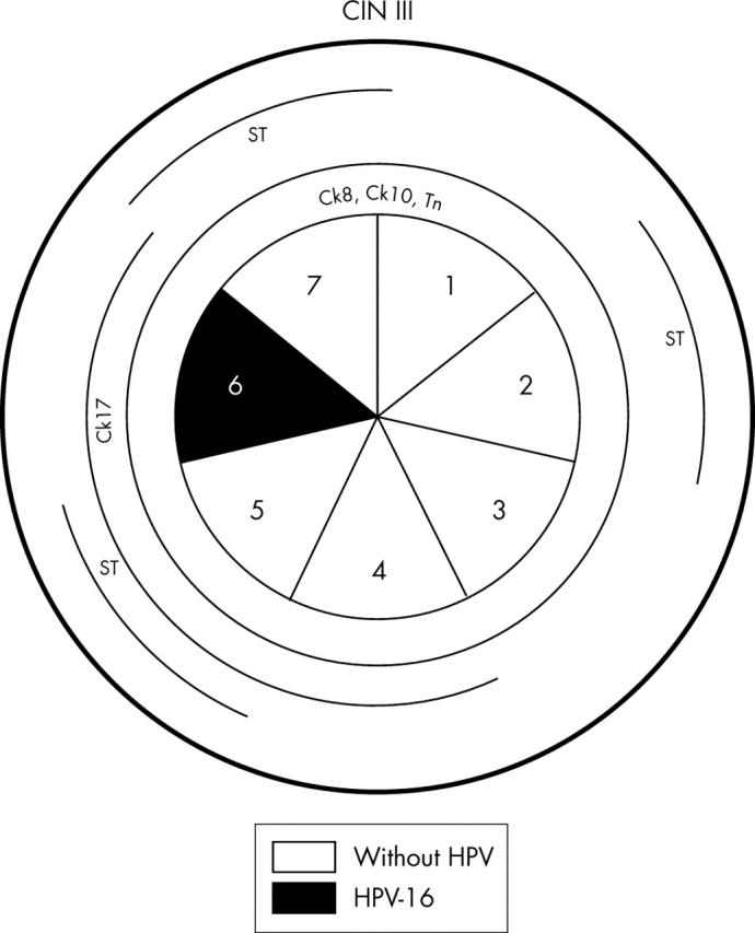
Schematic representation of case 4, including the absence or presence of human papillomavirus (HPV) and HPV types in the seven sections. Lesions of cervical intraepithelial neoplasia (CIN) III were observed in the whole specimen and are outlined in the outer circle. Different cytokeratins (Cks) and glycoprotein markers are outlined in the inner circles whenever their pattern of expression was abnormal.