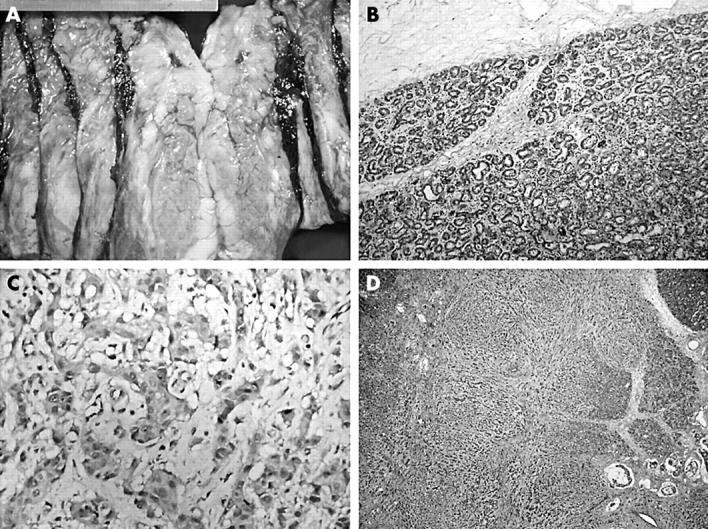
Figure 1.

(A) Gross photograph showing cream coloured tumour at the top (the central cystic haemorrhagic area is the result of a previous biopsy) merging with a lactating adenoma at the bottom. (B) The lactating adenoma is well circumscribed and characterised by ducts lined with vacuolated secretory cells. (C) High power magnification of the infiltrating adenocarcinoma demonstrating its high grade nature. (D) Low power magnification showing an invasive carcinoma displaying mixed morphology infiltrating between the lobules of the lactating adenoma.
