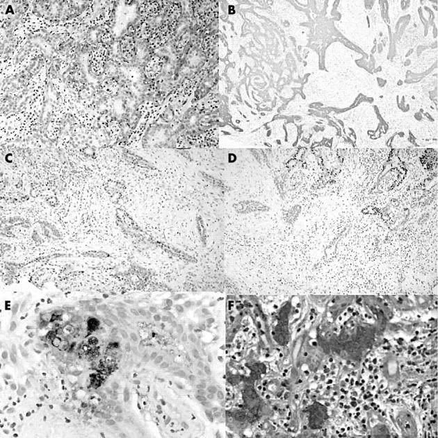
Figure 2.

Immunohistochemical staining for (A) muscle specific actin; (B) cytokeratin 7; (C and D) oestrogen and progesterone receptor, respectively (note the loss of immunoreactivity in the lower lefthand corner); and (E) gross cystic disease fluid protein. (F) Staining for periodic acid Schiff.
