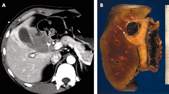
Figure 1.
(A) Abdominal computerised tomography image showing partial infiltration of the tumour into the gallbladder wall but no sign of invasion of the liver or portal vein. (B) Macroscopically, the gallbladder lumen is filled with blood and degenerative tissue, and the cut surface of the tumour has a nodular, well circumscribed, glistening appearance.

