
Figure 1.
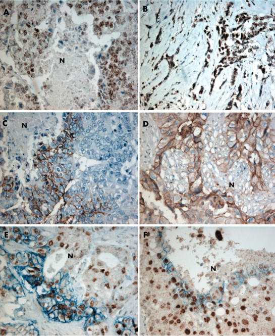
Immunohistochemical analysis of hypoxia inducible factor 1α (HIF-1α) and carbonic anhydrase IX (CA IX) in human breast cancer. (A) HIF-1α nuclear expression around necrotic (N) areas. (B) Diffuse nuclear HIF-1α expression, not related to necrosis. (C) CA IX membrane expression around necrotic areas. (D) Glucose transporter 1 (GLUT-1) membrane expression around necrotic areas. (E, F) Double staining revealing strong staining of HIF-1α and CA IX around necrotic areas (original magnification, ×200).