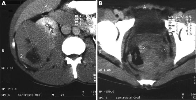
Figure 2.
(A) Abdominal computed tomography scan showing a solid tumour with hypodensities suggestive of necrosis in the hepatorenal space and retroperitoneal cavity, causing a compression effect against the right kidney. (B) Pelvic computed tomography scan showing the presence of a solid tumour at the rectum–sigmoid level, with hypodensities indicated by the numbers 1, 2, and 3.

