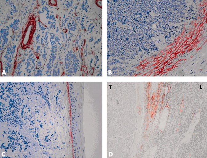
Figure 1.

(A) Strong tenascin-C (Tn-C) expression around the vascular structures within the tumour tissue. The tumour cells show no Tn-C expression. (B) Expression of Tn-C in the invasion fronts of the tumour, with flame-like extensions. (C) Moderate Tn-C staining in the dermo–epidermal junction of the normal skin. (D) Expression of Tn-C in metastatic lymph node. Septae show moderate to strong staining intensity, whereas normal lymphatic tissue (L) and metastatic tumour tissue (T) are negative. (A–D) Original magnification, ×200.
