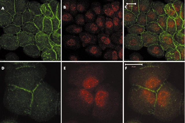
Figure 5.
Subcellular localisation of axin1 in MCF12A cells. Based on the known function of axin1, it is expected to be mainly localised in the cytoplasm. Confocal sections of MCF12A cells stained with (A and D) anti-axin1 and (B and E) anti-β catenin antibodies (C and F merged) show both nuclear and cytoplasmic axin1 staining, with a pronounced nuclear localisation, whereas β catenin is located mainly at the cell membrane (scale bars, 20 μm). The following antibodies were used: polyclonal anti-axin1 (1/500 dilution; Zymed, South San Francisco, California, USA), monoclonal anti-β catenin antibody (1/500 dilution; Signal Transduction Laboratories; Lexington, Kentucky, USA), secondary monoclonal and polyclonal antibodies (1/1000 dilution; Molecular Probes, Eugene, Oregon, USA). Other studies show a more cytoplasmic localisation for axin1. However, the subcellular localisation of axin1 appears to be cell type dependent.

