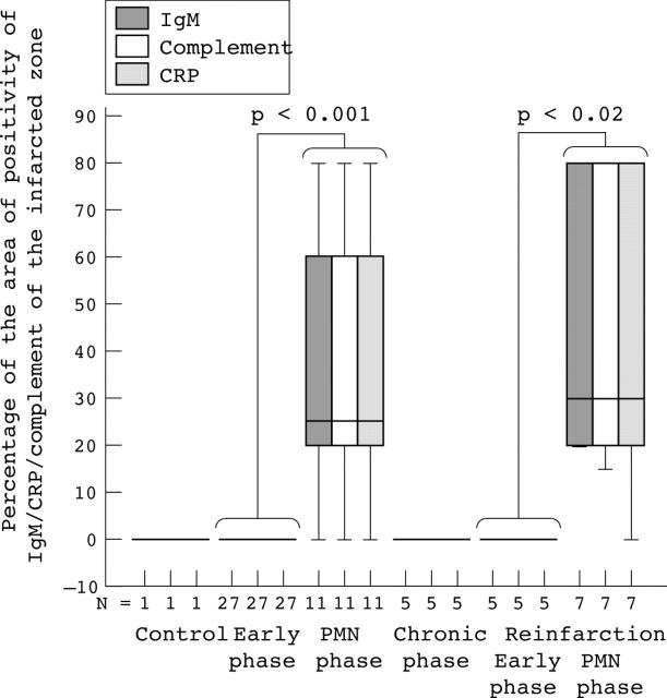
Figure 2.
Extent of IgM, complement, and C reactive protein (CRP) deposition in the infarcted myocardium. Box plot presentation of the percentage of IgM (grey bars), complement (white bars), and CRP (shaded bars) positive myocardium. For each patient, the percentage of positive surface area for the particular antibody in relation to the total area of the examined tissue was calculated. The error bars represent minimum and maximum values, whereas the boxes represent the lower and upper quartiles. The black lines within the boxes represent the medians (N, the number of patients examined). PMN, polymorphonuclear leucocyte.

