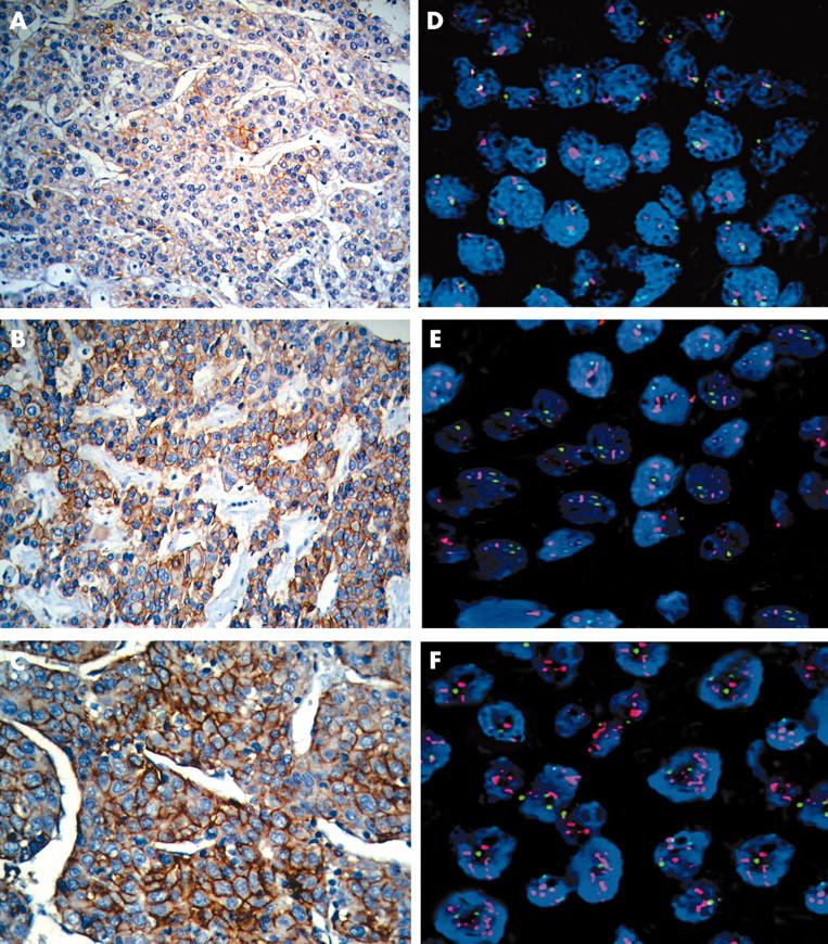
Figure 1.

(A–C) Membrane staining in neoplastic cells with the anti-HER-2/neu antibody. (D–F) Fluorescence in situ hybridisation results. Red and green signals represent HER-2/neu and chromosome 17 hybridisations, respectively, and the blue DAPI stain demonstrates the nuclei.
