
Figure 2.
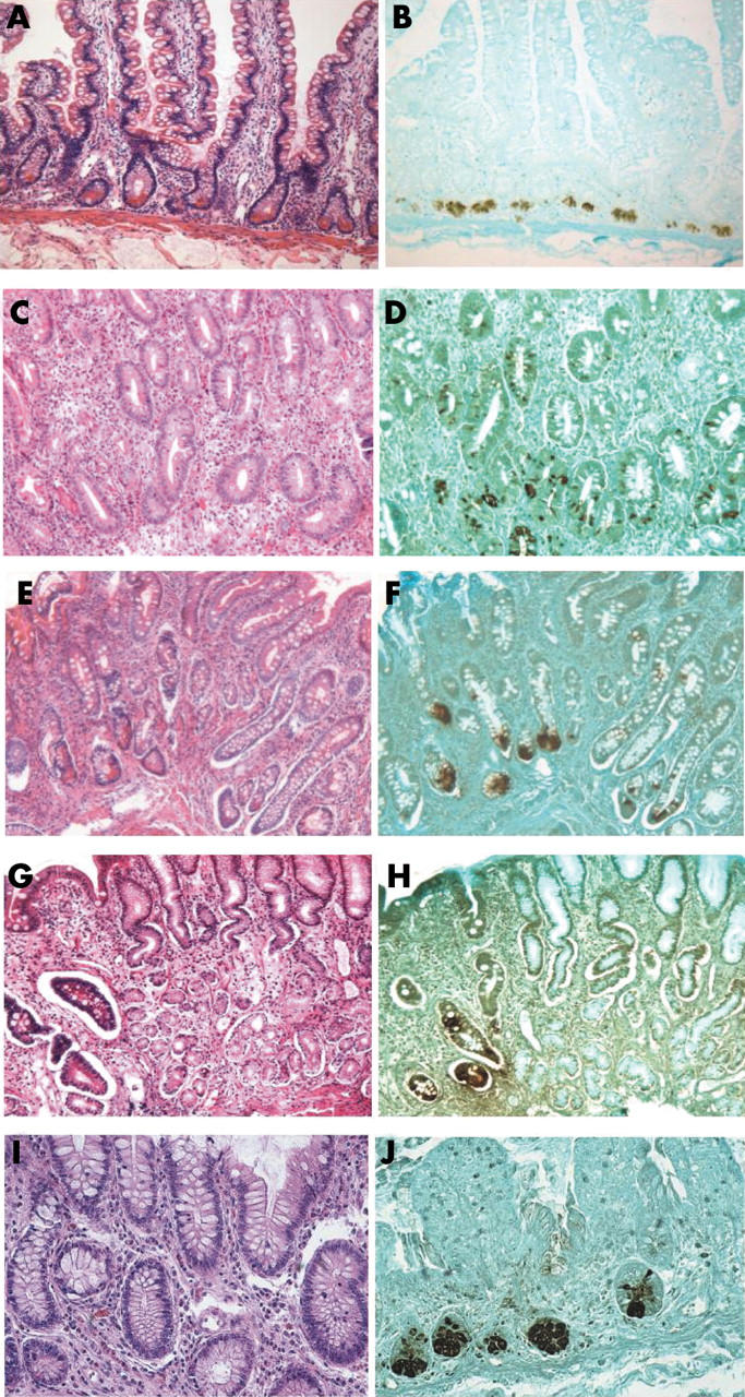
Detection of human defensin 5 (HD5) expression in gastrointestinal tissues by immunohistochemistry. Tissue sections were stained with the 8C8 monoclonal anti-HD5 antibody (B, D, F, H, and J), using a counterstain of light green. Parallel tissue sections were stained with haematoxylin and eosin (A, C, E, G, and I). The tissues were from the small intestine (A, B), Barrett’s oesophagus with goblet cell metaplasia (C, D), gastric intestinal metaplasia (GIM) with goblet cells (E, F), GIM at the border with normal gastric mucosa (G, H), and ileal mucosa from ileal pouch after colectomy with an ileal pouch–anal anastomosis (I, J). The pyramid shaped Paneth cells shown in (A) have characteristic eosinophilic secretory granules when viewed at higher power, but few or no eosinophilic granules are detectable in C, E, G, or H.