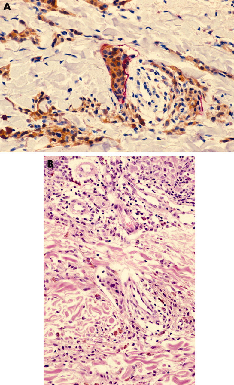
Figure 6.

(A) LYVE-1/S100 stained section showing a cluster of melanoma cells within a LYVE-1 positive peritumorous lymphatic vessel (LYVE-1 stains red; S100 stains brown; original magnification, ×60). (B) A tumour embolus is present but less apparent in a serial section stained with haematoxylin and eosin (original magnification, ×60).
