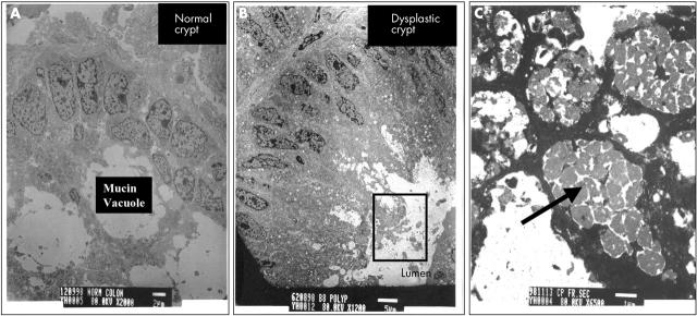
Figure 7.
Transmission electron microscopy micrographs of (A) a normal colonic crypt (scale bar, 2 μm) and (B) a dysplastic crypt (scale bar, 5 μm). The inset (C) shows a magnified view of the pigmented structures in the apical region of the dysplastic crypt, identified as “lipofuscin-like” material (scale bar, 1 μm).

