
Figure 1.
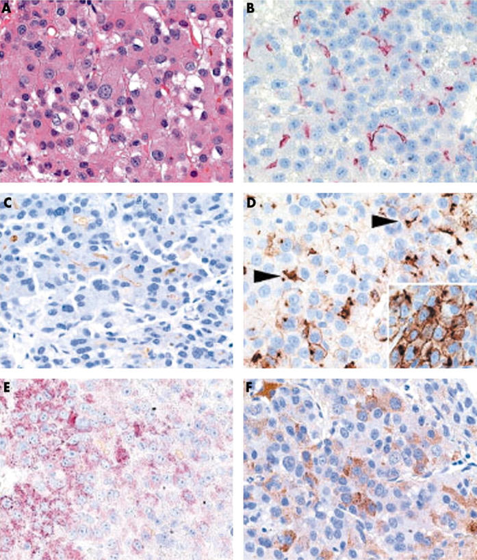
(A) A moderately differentiated hepatocellular carcinoma showing canalicular expression of (B) CD13 (aminopeptidase N), (C) p-CEA (antibody that crossreacts with biliary glycoprotein I), and (D, arrowheads) CD10, and cytoplasmic staining with (E) HepPar1 and (F) anti-AFP (α fetoprotein). (D, insert) Additional strong membranous expression was seen for CD10. (A) Haematoxylin and eosin; (B) anti-CD13, (C) anti-p-CEA, (D) anti-CD10, (E) HepPar1, and (F) anti-AFP, all with haematoxylin counterstain; original magnifications, ×400.