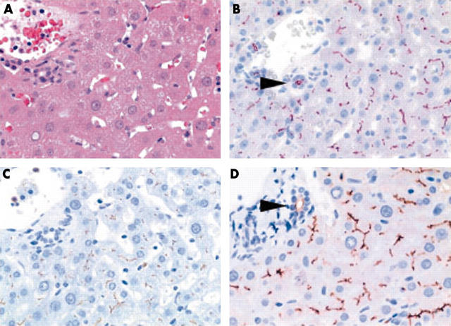
Figure 2.
(A) Non-neoplastic liver tissue showing canalicular immunostaining with (B) anti-CD13 (aminopeptidase N), (C) anti-p-CEA (antibody that crossreacts with biliary glycoprotein I), and (D) anti-CD10. Bile ducts express CD10 and CD13 at the apical membrane (B and D; arrowheads). (A) Haematoxylin and eosin; (B) anti-CD13, (C) anti-p-CEA, and (D) anti-CD10, all with haematoxylin counterstain; original magnifications, ×400.

