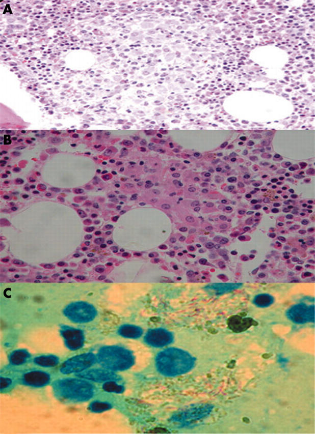
Figure 1.

(A) Bone marrow aspirates showing many histiocytes with needle-like inclusions in the cytoplasm in patient 1. (B) Bone marrow biopsy showing granulomatous lesions with aggregates of histiocytes in patient 1. (C) Lymph node aspirate showing acid fast bacilli in foamy histiocytes in patient 2.
