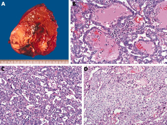
Figure 1.

(A) Macroscopically, the nodular S8 tumour has a fibrous capsule. The cut surface of the tumour is well circumscribed, expanded, and yellow/white to dark red in colour. (B) The tumour cells have abundant eosinophilic granular cytoplasm and round nuclei with moderate variations in size and shape. The irregular trabecular structures are filled with a bloody/serous or bloody fluid without mucin production and desmoplastic tissue. The typical trabecular pattern was not seen (haematoxylin and eosin (H&E) stain, original magnification, ×200). (C) Small tubular or acinar-like patterns are also visible (H&E stain; original magnification, ×200). (D) Solid structures are seen in a small portion of the tumour (H&E stain; original magnification, ×200).
