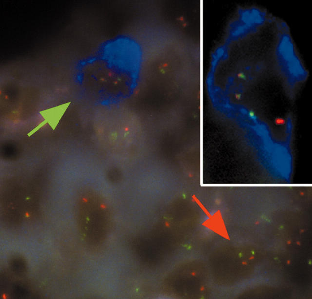
Figure 1.
FICTION analysis of a mantle cell lymphoma bone marrow biopsy prepared using EDTA based Osteosoft. Bone marrow sections were immunostained with an anti-CD20 antibody and AlexaFluor 350 conjugated antibodies and streptavidin. Dual fusion fluorescence in situ hybridisation (FISH) probes for the detection of t(11;14)(13q;32q) were then applied. The blue immunostaining indicates CD20 positive cells and the red and green signals indicate the CCND1 and IGH loci, respectively. Normal cells show two red and two green FISH signals (red arrow), whereas abnormal cells harbouring a CCND1/IGH translocation show one red, one green, and two fused FISH signals (green arrow and inset).

