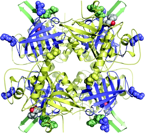
Figure 3. The biologically active tetrameric form of hDPPI in complex with the covalently bound inhibitor Gly-Phe-CHN2.
The monomers are located at the four corners of the tetramer with the exclusion domains shown in blue and the papain-like domains shown in pale yellow. The active sites can be seen on the outside of the tetramer by the location of the inhibitors which are shown with atoms as grey spheres. The structural elements close to the inhibitor-binding site, the N-linked carbohydrate at Asn5 and the β-hairpin from Lys82 to Tyr93 are shown in pale green. The other N-linked carbohydrates are shown as spheres in the same colour as the domains to which they are linked.

