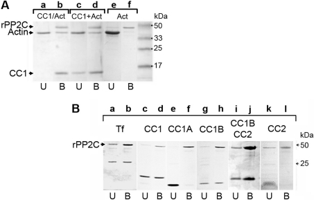
Figure 4. The CC1–G-actin complex binds to PP2C through CC1B but not through CC1A.
(A) Immobilized rPP2C on Ni-NTA was incubated with either the preformed CC1–G-actin complex (lanes a and b) or sequentially with CC1 then G-actin (lanes c and d) or with G-actin alone (lanes e and f) for 1 h at 4 °C. After extensive washings, Ni-NTA resin-bound proteins were eluted in SDS sample buffer and subjected to SDS/PAGE and Coomassie Blue staining. U indicates the total unbound fraction and B indicates the total bound fraction. Arrowheads on the left indicate the positions of G-actin, CC1 and rPP2C. (B) Immobilized rPP2C was incubated with toxofilin (Tf; lanes a and b), CC1 (lanes c and d), CC1A (lanes e and f), CC1B (lanes g and h), CC1BCC2 (lanes i and j) or CC2 (lanes k and l). Samples were treated as described for Figure 4(A). The arrowhead on the left indicates the position of rPP2C.

