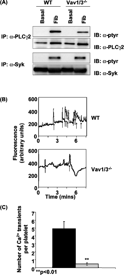
Figure 2. Activation of PLCγ2 by αIIbβ3 is impaired in Vav1/3−/− platelets.
(A) Washed wild-type (WT) and Vav1/3−/− platelets were incubated over fibrinogen-coated Petri dishes in the presence of 2 units/ml apyrase and 10 μM indomethacin for 45 min at 37 °C. Basal (non-adherent) and fibrinogen-stimulated (adherent; Fib) cells were lysed in Nonidet P40 lysis buffer, and PLCγ2 and Syk were immunoprecipitated (IP). Immunoprecipitates were separated by SDS/PAGE and were Western-blotted (IB) with anti-phosphotyrosine monoclonal antibody 4G10 (α-ptyr). Membranes were subsequently stripped and reprobed with anti-PLCγ2 (α-PLCγ2) and anti-Syk (α-Syk) antibodies respectively. (B) Washed wild-type (WT) and Vav1/3−/− platelets were labelled with the Ca2+ reporter dye Oregon Green BAPTA-1/AM and incubated over fibrinogen-coated coverslips in the presence of 2 units/ml apyrase and 10 μM indomethacin and imaged in real time using the FITC channel on a fluorescent microscope. The fluorescence intensity of a representative platelet is shown. Spikes in fluorescence indicative of transient cytoplasmic Ca2+ elevation are indicated by arrows. (C) The number of Ca2+ transients in 30 platelets chosen at random from wild-type (black bar) and Vav1/3−/− (grey bar) mice were counted. Results are means±S.E.M. and are representative of three experiments.

