
Figure 2.
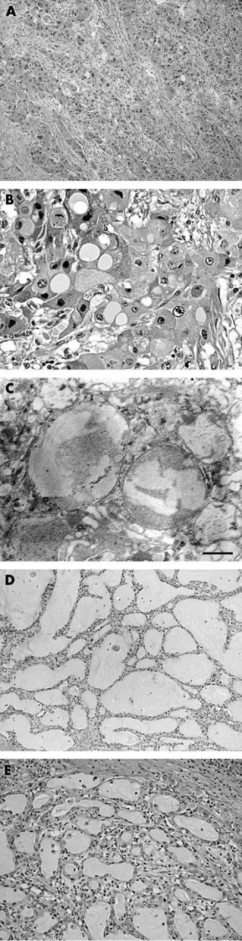
Histology of the tumour. (A) In fibrolamellar carcinoma, the tumour nests, consisting of large, eosinophilic neoplastic hepatocytes, are embedded in a fibrous stroma arranged in a lamellar fashion. (B) Some of the tumour cells contain sharply demarcated, pale staining, round structures (pale bodies) in their cytoplasm. (C) Electron microscopic observation of the pale bodies reveals bundles of fine fibrillar material present in membrane bound structures. The scale bar indicates 500 nm. (D) The cholangiocarcinoma (CC) tumour consists of irregular glandular structures surrounded by a fibrous stroma. (E) Higher magnification of an area of CC. The glandular lumens are filled with mucin.