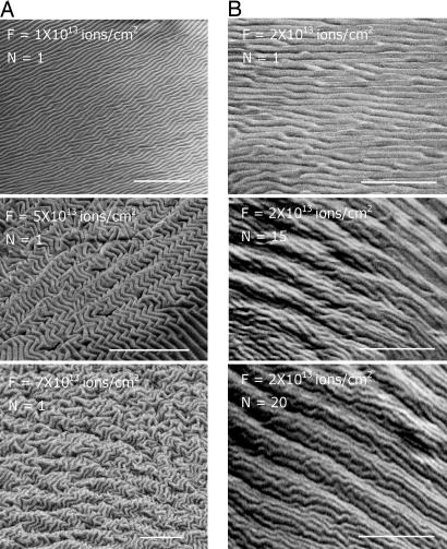
Fig. 2.
The surface morphology of the wrinkle patterns induced by FIB depends primarily on the applied ion fluence. (A) SEM images of the surface morphology of the PDMS exposed to FIB in a single scan mode (N = 1) with various ion beam fluences, F (ions per cm2). (B) SEM images of the surface morphology created by N scans each with fluence, F = 2.0 × 10¹³ ions per cm2. (Top) SEM image shows the surface morphology after the first scan. AFM examination of the surface shows that the surface has a wavelength of ≈470 nm. (Middle and Bottom) SEM images of the surface after N = 15 (Middle) and 20 (Bottom) scans (with accumulated fluence, N × F, of 3.0 × 10¹⁴ and 4.0 × 10¹⁴ ions per cm2, respectively) reveals complex patterns of the surface with a hierarchical nature. (Scale bars: 10 μm.)

