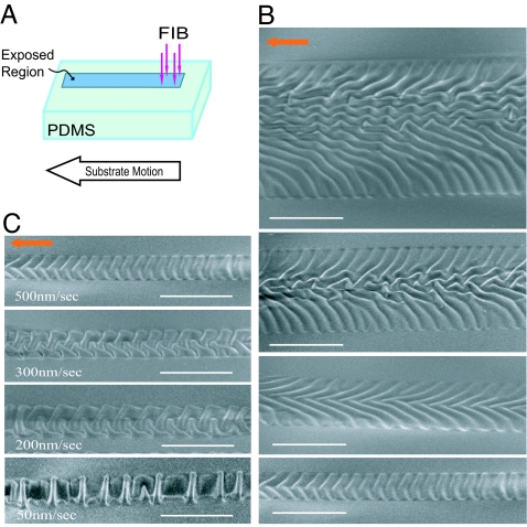
Fig. 4.
Various morphologies of the wrinkled skins induced by controlling the relative motion of the ion beam and PDMS. (A) By moving the PDMS stage during the FIB irradiation, long channel patterns can be produced. (B) SEM images of the regions of the PDMS exposed to FIB showing various surface morphologies created by changing the width from 20 μm (top image) to 4 μm (bottom image). The PDMS stage has a constant velocity of 500 nm/s and the beam current is 1 pA. The ion fluence varies from 7.2 × 10¹³ to 1.8 × 10¹⁵ ions per cm2 (top to bottom images). (C) Surface morphologies created by varying the moving speed of the PDMS stage from 50 to 500 nm/s, while the width of the exposed area is kept constant as 4 μm. The beam current is constant at 1 pA such that the fluence varies from 1.8 × 10¹⁵ to 1.8 × 10¹⁶ ions per cm2. Note that the overall shape of the patterns also can be controlled as shown in Fig. 1D. (Scale bars: 10 μm.)

