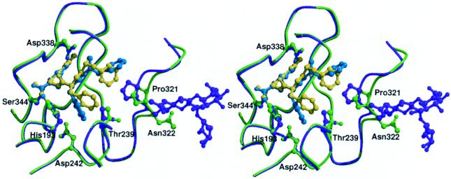
Figure 3.
Comparison of the inhibitor-binding mode in Gla-domainless (purple) and sTF-complexed fVIIa (light green). The FFR inhibitor in the GD-fVIIa and fVIIasTF structures is colored cyan and yellow, respectively. The carbohydrate groups attached to Asn-322 {175} in the GD-fVIIa structure are also shown.

